|
Archive
Blog
Cast
Forum
RSS
Books!
Poll Results
About
Search
Fan Art
Podcast
More Stuff
Random
Support on Patreon |
|
New comics Mon-Fri; reruns Sat-Sun
|
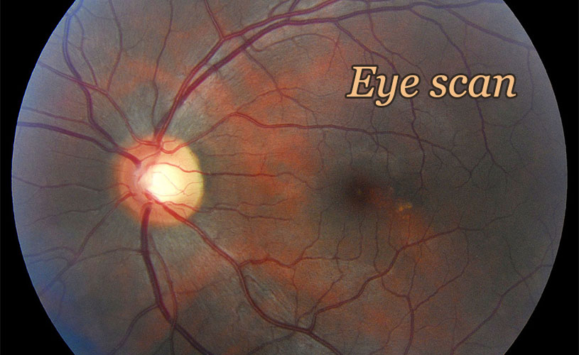
1 {photo of a retina}
1 Caption: Eye scan
|
First (1) | Previous (3330) | Next (3332) || Latest Rerun (2580) |
Latest New (5175) First 5 | Previous 5 | Next 5 | Latest 5 Annotations theme: First | Previous | Next | Latest || First 5 | Previous 5 | Next 5 | Latest 5 This strip's permanent URL: http://www.irregularwebcomic.net/3331.html
Annotations off: turn on
Annotations on: turn off
|
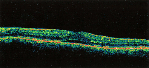 OCT scan of my left retina, back in January 2014, showing blistering of the retina. |
I had noticed my vision improving slowly over the past few months, and now it seems to be the same as it was before I noticed the problem initially. The ophthalmologist took another OCT (optical coherence tomography) scan of each of my eyes. The blistering that was present in January has vanished, leaving my retina back to normal, with the expected indent where the fovea is. This is excellent news, from my point of view (pun intended).
The ophthalmologist said the average recurrence rate of central serous retinopathy is about 2.3 cases per patient. Because of the spread of the distribution, this means that it is most likely not to recur in my case. But if it does recur, then it might recur 4 or 5 times before vanishing as mysteriously as it came. There seem to be a surprising number of medical conditions for which modern medicine still hasn't worked out what causes them.
Optical coherence tomography is a cool medical imaging technique, which allows the taking of these sorts of retinal images. Like x-ray computed tomography (commonly known as CT scans), OCT produces an image which is a cross-section of the body being imaged, rather than a face-on image such as would be made with a conventional camera. How does it do this?
The retina is made of several partially transparent layers, which allow the incoming light to penetrate through the overlying tissues to the light-sensitive layer of rod and cone cells. These layers reflect different fractions of the incoming light. If you can measure the fraction of light reflected at different depths, you could build up a cross-sectional image of the layers within the retina. This is exactly how OCT works.
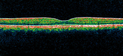 OCT scan of my left retina, May 2014, showing complete healing of the retina. |
The light reflected back from the movable mirror (B) is recombined with the light reflected back from the back of the patient's eyeball. Now, remember that light has wave properties. If the distance from the semi-reflective mirror (A) to the patient's retina is exactly the same as the distance from the semi-reflective mirror (A) to the movable mirror (B), then the waves of the two light beams will interere constructively, reinforcing one another, and you will produce a bright light. If the path lengths are different, then because of the mix of wavelengths in the light, many of the wavelengths will interfere destructively, the result being a much weaker light intensity.[1] In this way, the interferometer preferentially picks out the reflection from the layer of the retina that is exactly the same distance from the semi-reflective mirror (A) as is the movable mirror (B). Now, by scanning the movable mirror (b) back and forth, thus adjusting the path length of the light, you can scan in depth within the retina.
This depth scan gives you different intensities of light, depending on how reflective the layer of the retina is at each specific depth. And so you can build up a picture of the structure within the retina at each spot. Then the transverse scan of the light beam gives you a slice through the retina, resulting in the sort of image you see here, as the output of the OCT scan.
Pretty cool, huh?
|
LEGO® is a registered trademark of the LEGO Group of companies,
which does not sponsor, authorise, or endorse this site. This material is presented in accordance with the LEGO® Fair Play Guidelines. |